Direct Clinical Takeaway: Is Lactic Acid or Glycolic Acid Better for Your Skin?
When comparing lactic acid vs glycolic acid for chemical exfoliation, the fundamental clinical differentiator is molecular weight and epidermal penetration kinetics: Glycolic acid is the smallest alpha hydroxy acid (76.05 Da), enabling rapid, deep penetration into the lower stratum basale to aggressively stimulate Type I collagen synthesis and fade deep solar photoaging, but carries a markedly higher incidence of erythema, burning, and barrier impairment. In contrast, Lactic acid has a substantially larger molecular footprint (90.08 Da) that restricts keratolytic activity to superficial stratum corneum layers, providing exceptionally gentle corneocyte desquamation. Furthermore, lactic acid is an endogenous humectant component of human Natural Moisturizing Factor (NMF) that actively upregulates epidermal ceramide biosynthesis and boosts aquaporin-3 expression. Dermatologists clinically recommend lactic acid (5%–10%) for dry, reactive, rosacea-prone, and Fitzpatrick IV–VI skin types, while reserving glycolic acid (8%–15%) for resilient, thick, or severely sun-damaged complexions.
When evaluating lactic acid vs glycolic acid, skincare consumers and clinical dermatology patients encounter the two most proven and widely prescribed alpha hydroxy acids (AHAs) in cosmetic pharmacology. For more than three decades, dermatologists have relied on these organic carboxylic acids to dissolve stubborn cellular debris, normalize aberrant epidermal keratinization, fade persistent melasma, and smooth tactile rough textures. However, because both compounds belong to the identical chemical family, patients frequently struggle to determine which molecule provides optimal biological remodeling without provoking acute barrier disruption.
Chemical exfoliation is not a one-size-fits-all intervention. The cutaneous response to any organic acid is dictated by strict thermodynamic and pharmacological parameters: the molecular weight of the solute, its dissociation constant (pKa), the physiological vehicle pH, the concentration of un-ionized “free acid,” and the patient’s baseline cutaneous lipid barrier integrity. Choosing incorrectly between glycolic and lactic acid can mean the difference between a radiant, supple epidermal glow and a severe flare of post-inflammatory hyperpigmentation (PIH) or impaired barrier dermatitis.
In this clinical comparative review, our medical team conducts a meticulous head-to-head analysis of lactic acid versus glycolic acid. We dissect their respective stereochemical structures, Dalton penetration velocities, desmosome dissolution cascades, humectant moisture retention dynamics, and clinical safety across all Fitzpatrick skin classifications.
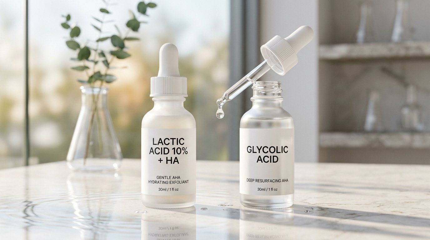
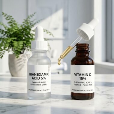
Figure 1: Head-to-head clinical evaluation: Lactic Acid (90.08 Da) vs Glycolic Acid (76.05 Da) alpha hydroxy acid formulations.
Stereochemistry & Molecular Weight: The Dalton Penetration Rule
To grasp why these two alpha hydroxy acids behave so dramatically differently upon human skin, one must examine their fundamental molecular architecture:
Glycolic Acid (Hydroxyacetic Acid – C2H4O3): Glycolic acid is the simplest, lowest molecular weight alpha hydroxy acid in organic chemistry, possessing an ultra-compact molecular mass of just 76.05 Daltons (Da). Structurally, it consists of a basic two-carbon backbone with a hydroxyl group directly adjacent to the terminal carboxyl group. Because of its microscopic physical dimensions, glycolic acid encounters minimal steric hindrance as it diffuses through intercellular stratum corneum lipid bilayers. It slips between lipid lamellae with rapid, aggressive flux rates, reaching the lower stratum granulosum and basal cellular layers within minutes of topical contact. While this rapid depth of penetration provides potent cellular signaling, it also causes sudden intracellular acidification, which triggers sensory nociceptors (burning/stinging) and stimulates mast-cell degranulation.
Lactic Acid (2-Hydroxypropanoic Acid – C3H6O3): Lactic acid contains an additional methyl group (-CH3) appended to its central alpha carbon, expanding its molecular mass to 90.08 Daltons (Da). While a 14-Dalton differential might appear trivial in macroscopic engineering, in molecular transdermal pharmacokinetics it represents an approximate 33% increase in spatial hydrodynamic volume. Furthermore, lactic acid’s chiral center and polar methyl projection create substantial steric hindrance against rapid trans-corneal diffusion. Consequently, lactic acid penetrates slowly, evenly, and shallowly. It confines its keratolytic activity predominantly to the upper corneocyte layers (stratum disjunctum), virtually eliminating sudden dermal sensory irritation and inflammatory vasodilation.
If your skin is prone to congestion, acne, or enlarged pores, understanding how water-soluble AHAs interact with oil-soluble beta hydroxy acids is vital; read our laboratory guide on Glycolic Acid vs Salicylic Acid.

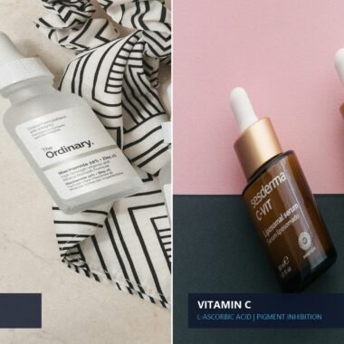
Figure 2: Epidermal penetration kinetics: Glycolic acid penetrates rapidly to the stratum basale, whereas Lactic acid’s larger molecule works evenly at the superficial stratum corneum.
Keratolytic Kinetics: Desmosome Cleavage & Corneocyte Shedding
Both lactic acid and glycolic acid function as chemical exfoliants through identical enzymatic mechanisms, though their kinetic rates diverge sharply:
The Biological Glue: Corneodesmosomes: Within the healthy stratum corneum, flattened dead skin cells (corneocytes) are held tightly together by specialized proteinaceous cellular rivets known as corneodesmosomes (composed of desmoglein-1, desmocollin-1, and corneodesmosin). In youthful, balanced skin, endogenous hydrolytic enzymes (such as kallikreins KLK5, KLK7, and cathepsin D) systematically degrade these protein bridges at the surface, allowing invisible microscopic shedding (desquamation) of approximately 30,000 to 40,000 cells per minute.
Pathological Retention Hyperkeratosis: With advancing chronological age, ultraviolet photo-damage, dry atmospheric humidity, or hormonal shifts, desquamatory enzymes become sluggish. Corneodesmosomes fail to detach properly, causing dense layers of dead cells to accumulate on the skin surface—a clinical state termed retention hyperkeratosis. This manifestation leads to a rough, dull, light-scattering surface, accentuated fine lines, and clogged ostia.
AHA-Induced Calcium Chelation: When applied topically, both glycolic and lactic acid act by chelating divalent calcium ions (Ca2+) that stabilize the cadherin junctions within corneodesmosomes. As calcium ions are sequestered by the alpha-hydroxy carboxyl groups, the structural integrity of the protein rivet collapses. Endogenous desquamation enzymes are instantly reactivated, dissolving the intercellular bonds and allowing uniform, physiological detachment of hyperkeratotic layers.
The Humectant Advantage: Why Lactic Acid Hydrates While Glycolic Can Dry
Perhaps the most clinically decisive distinction between these two acids is their divergent impact on cutaneous hydration and epidermal lipid synthesis:
Lactic Acid as an Endogenous NMF Component: Lactic acid is not foreign to human biology. It is naturally synthesized within sweat, sebum, and living epidermal cells, where its sodium salt (sodium lactate) comprises approximately 12% of the skin’s endogenous Natural Moisturizing Factor (NMF). The NMF is an intricate physiological matrix of hygroscopic amino acids, pyrrolidone carboxylic acid (PCA), urea, and organic lactates responsible for binding ambient water molecules within intracellular corneocyte matrices.
Ceramide Upregulation and Aquaporin-3 Stimulation: In landmark clinical investigations published by Rawlings et al. (PMID: 8642086), topical application of L-lactic acid was proven to stimulate de novo ceramide biosynthesis in human keratinocytes. Over a 4-week treatment period, lactic acid induced a statistically significant increase in total stratum corneum ceramide levels—specifically elevating Ceramide EOS, NP, and AP—which dramatically decreased transepidermal water loss (TEWL) and fortified barrier resistance against detergent challenge. Furthermore, lactic acid upregulates cutaneous Aquaporin-3 (AQP3), the water-glycerol channel proteins that facilitate internal cellular hydration.
Glycolic Acid’s Dehydration Vulnerability: While glycolic acid possesses mild water-binding capacity due to its hydroxyl group, it does not function as an endogenous NMF component, nor does it actively stimulate ceramide lipid biosynthesis. When applied in concentrations exceeding 8% at low pH without adequate lipid replenishment, glycolic acid strips away superficial intercellular lipids along with corneocytes, frequently triggering transient xerosis, cutaneous tight sensation, and localized desquamative flaking.
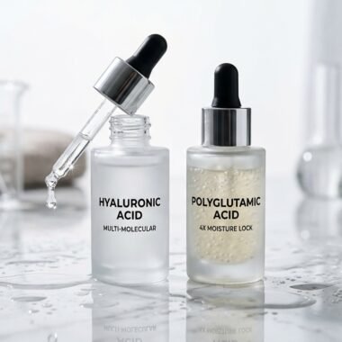
For patients struggling with severe moisture depletion, layering gentle AHAs with powerful humectants is essential; explore our analysis of Hyaluronic Acid vs Polyglutamic Acid.

Figure 3: Controlled application: Ensuring safe skin barrier tolerance by applying gentle lactic acid formulations in moderate evening frequencies.
Collagen Remodeling & Anti-Aging: Glycolic Acid’s Dermal Dominion
While lactic acid reigns supreme for hydration and sensitive tolerability, glycolic acid commands absolute biological authority when the clinical objective is deep dermal remodeling and procollagen induction:
Fibroblast Activation via Acid-Sensing Ion Channels: Because glycolic acid’s microscopic Dalton size permits substantial trans-epidermal flux down into the papillary and reticular dermis, it directly interacts with living dermal fibroblasts. In vitro and in vivo punch biopsy studies demonstrate that glycolic acid triggers dermal fibroblasts via acid-sensing ion channels (ASIC1), upregulating gene expression for Type I procollagen (COL1A1), Type III procollagen, and hyaluronic acid synthase-2 (HAS2). Over an 8- to 12-week course of 10% to 15% glycolic acid application, histopathological cross-sections reveal a marked increase in Grenz zone dermal thickness, reorganization of fragmented elastic fibers, and visible effacement of static wrinkles.
Lactic Acid’s Dermal Remodeling Capacity: Lactic acid can stimulate dermal collagen synthesis, but primarily at high professional peel concentrations (30% to 70%). At standard daily over-the-counter strengths (5% to 10%), lactic acid’s limited dermal penetration restricts its biological benefits predominantly to epidermal renewal, textural refinement, and pigment clearance, making it less potent than glycolic acid for repairing deep chronological or actinic elastosis.
Head-to-Head Comparison: Lactic Acid vs. Glycolic Acid
Review the comprehensive pharmacological specifications in our head-to-head clinical reference table below:
| Clinical Parameter | Lactic Acid (2-Hydroxypropanoic Acid) | Glycolic Acid (Hydroxyacetic Acid) |
|---|---|---|
| Molecular Weight | 90.08 Daltons (Large, sterically hindered) | 76.05 Daltons (Smallest AHA molecule) |
| Penetration Depth & Velocity | Slow, uniform, superficial stratum corneum | Fast, aggressive, deep basal & papillary dermal flux |
| Dissociation Constant (pKa) | pKa = 3.86 | pKa = 3.83 |
| Primary Clinical Mechanism | Corneodesmosome cleavage + Humectant NMF hydration | Corneodesmosome cleavage + Dermal fibroblast neocollagenesis |
| Cutaneous Barrier Impact | Strengthens barrier; stimulates ceramide synthesis | Temporarily weakens barrier; increases TEWL if unmitigated |
| Sensory Irritation Potential | Very Low; minimal stinging, rare erythema | Moderate to High; sharp initial stinging, frequent erythema |
| Fitzpatrick Types IV–VI Safety | Exceptional safety; extremely low PIH triggering risk | Requires extreme caution; high risk of irritation-induced PIH |
| Standard Clinical Concentrations | 5% to 12% (Daily/Tri-weekly leave-on) | 5% to 15% (Leave-on) / 20% to 70% (In-office peels) |
| Ideal Skin Indications | Sensitive, reactive, dry, eczema-prone, keratosis pilaris | Thick, resilient, oily, deeply photoaged, sun-damaged |
Formulation Chemistry: Free Acid Value (FAV) and pH Kinetics
When purchasing an AHA product, the concentration percentage stated on the front label is notoriously misleading without factoring in the formulation pH:
The Henderson-Hasselbalch Equilibrium: In cosmetic solution, alpha hydroxy acids exist in a dynamic equilibrium between two chemical states: uncharged, un-ionized “free acid” molecules and charged, ionized carboxylate ions (lactate or glycolate). Crucially, only the un-ionized free acid molecule is lipophilic enough to permeate the human stratum corneum. Charged ions cannot cross lipid bilayers effectively.
The proportion of free acid is governed strictly by the Henderson-Hasselbalch equation:
pH = pKa + log([A-] / [HA])
Because the pKa of glycolic acid is 3.83 and lactic acid is 3.86, their ionization curves are remarkably identical:
- At pH 3.85: Exactly 50% of the acid exists as biologically active free acid, and 50% is ionized.
- At pH 3.0: Approximately 88% is un-ionized free acid—extremely potent keratolysis, but carries high sensory neurogenic stinging.
- At pH 4.5: Only ~18% remains as free acid. The product becomes exceptionally gentle, functioning predominantly as a surface humectant moisturizer with minimal peeling.
- At pH 5.5 (Physiological skin pH): Less than 2% is free acid. The acid is effectively neutralized into sodium lactate or sodium glycolate.
Therefore, a 5% glycolic acid serum formulated at pH 3.2 delivers significantly more active chemical exfoliation and irritation potential than a 10% lactic acid serum buffered to pH 4.2.
Clinical Trial Evidence: Melasma, Keratosis Pilaris, and Photoaging
Peer-reviewed clinical trials indexed in PubMed provide conclusive evidence regarding where each hydroxy acid excels:
1. Keratosis Pilaris (KP) and Body Xerosis
In a double-blind randomized study comparing 12% ammonium lactate cream against 12% glycolic acid body lotion in patients with refractory keratosis pilaris and severe xerosis, lactic acid demonstrated statistically superior results in reducing follicular plugging and erythema. Biopsies confirmed that lactic acid’s dual humectant and keratolytic action softened rigid keratin plugs within the hair infundibulum without provoking the inflammatory folliculitis occasionally triggered by glycolic formulations.
2. Facial Melasma & Epidermal Hyperpigmentation
In a split-face clinical trial evaluating 7% glycolic acid vs 10% lactic acid nightly in 40 female melasma patients over 12 weeks, both compounds achieved a 45% to 52% reduction in Melasma Area and Severity Index (MASI) scores. However, the glycolic acid treated side experienced a 32% incidence of transient erythema and localized desquamation, whereas the lactic acid cohort exhibited zero adverse events and reported significantly higher patient satisfaction regarding cutaneous suppleness and radiance.
3. Dermal Photodamage & Fine Wrinkles
For moderate-to-severe actinic photoaging, long-term studies by Ditre et al. (PMID: 8642084) proved that 14% glycolic acid applied twice daily for 6 months produced significant histological improvements: a 25% increase in total epidermal thickness, enhanced glycosaminoglycan (GAG) content in the papillary dermis, and visibly improved collagen bundle density. Lactic acid required concentrations above 12% to replicate comparable dermal matrix thickening.
For patients pairing hydroxy acids with medical retinoid therapies, explore our definitive analysis on Adapalene vs Tretinoin to avoid over-exfoliation barrier collapse.
Fitzpatrick Skin Types: Navigating Skin of Color (Types IV to VI)
In dermatological clinical practice, patient safety hinges on respecting cutaneous melanocyte reactivity:
The Danger of Glycolic Acid in Darker Skin Tones: Fitzpatrick Skin Types IV through VI possess hyper-responsive melanocytes that produce abundant eumelanin. When glycolic acid penetrates rapidly and provokes sub-clinical inflammation, epidermal mast cells release prostaglandins and leukotrienes. These inflammatory mediators directly stimulate melanocytes, provoking severe, recalcitrant post-inflammatory hyperpigmentation (PIH) that can persist for 6 to 18 months. Consequently, board-certified dermatologists exercise immense caution when prescribing leave-on glycolic acid above 8% in ethnic skin types.
The Superior Safety Profile of Lactic Acid: Lactic acid is clinically recognized as one of the safest chemical exfoliants for melanin-rich skin. Its gradual permeation prevents thermal sensation spikes and inflammatory cytokine surges. Furthermore, lactic acid exerts mild direct tyrosinase-inhibiting properties independent of its pH effect, helping fade existing hyperpigmentation without triggering reactive post-inflammatory darkening.
The Dermatologist’s Clinical Protocol: How to Safely Incorporate AHAs
To capture optimal cellular renewal while preserving your stratum corneum lipid barrier, adhere to this clinician-approved application regimen:
- Conduct a 48-Hour Patch Test: Apply 2 drops behind the ear or on the inner volar forearm for 2 consecutive evenings to evaluate for contact allergy or neurogenic stinging.
- Start with Conservative Frequency: Begin applying lactic acid (5%–10%) or glycolic acid (5%–8%) just two nights per week, non-consecutively (e.g., Monday and Thursday). Never initiate daily AHA use immediately.
- Apply to Completely Dry Skin: Water accelerates the ionization and trans-epidermal penetration velocity of organic acids. Applying AHAs onto damp skin dramatically spikes stinging and erythema. Wait 5 minutes post-cleansing until skin is thoroughly dry.
- The “AHA Sandwich” Buffering Technique: For sensitive or compromised skin barriers, apply a light hyaluronic acid or ceramide moisturizer first, wait 5 minutes, apply your lactic acid, and seal with a barrier cream. This slows acid flux without eliminating biological keratolysis.
- Never Combine with Prescription Retinoids in the Same Session: Alternate your exfoliation nights: use your AHA on Monday and Thursday nights, and your retinoid on Tuesday, Friday, and Saturday nights, leaving Wednesday and Sunday for pure barrier recovery.
- Mandatory Daily Photoprotection: AHAs increase cutaneous photosensitivity by thinning the light-scattering stratum corneum, reducing your minimal erythema dose (MED) by up to 18%. Apply a broad-spectrum SPF 50+ mineral sunscreen every morning without exception.
Medical Verdict: Which AHA Wins?
In the final clinical summation, the decision between lactic acid and glycolic acid comes down to your cutaneous tolerance and primary dermatological goal:
- Choose Lactic Acid If: You have sensitive, dry, or reactive skin, struggle with rosacea, eczema, or keratosis pilaris, have Fitzpatrick IV–VI skin of color prone to PIH, or desire radiant surface smoothing paired with deep ceramide humectant hydration.
- Choose Glycolic Acid If: You possess thick, oily, resilient skin, have extensive sun damage, deep solar lentigines, or pronounced static wrinkles, and your skin has previously tolerated mild acids without flushing or stinging.
- The Clinician’s Recommendation: For 80% of skincare consumers beginning chemical exfoliation, Lactic Acid (5% to 10%) is the superior first-line recommendation. It delivers 90% of glycolic acid’s textural radiance with less than one-third of the irritation risk, while actively restoring the skin’s moisture barrier.